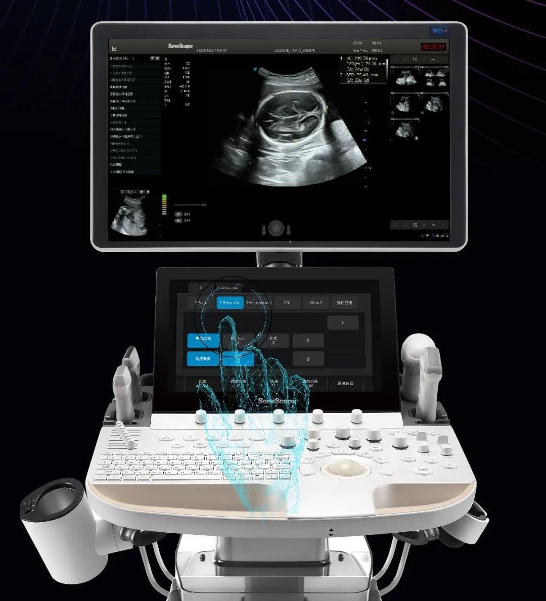
Extraordinary Image Technologies
With the outstanding clear view provided by SonoScape’s advanced imaging technologies and optimized transducers, you can make clinical decisions with greater confidence
-
μ-Scan
The new generation μ-Scan imaging technology greatly improves the visibility of organs and lesions. The high-definition contrast resolution will suppress speckle artifacts while maintaining real tissue architecture.
-
SR Flow
As a new innovative technology, SR Flow improves the capability of detecting low velocity flow signals. It also improves on spatial resolution and overcomes overflow to present users with real hemodynamic information
-
WideScan
With WideScan, the ultrasound image can be enlarged when performing a real time scan by using linear, convex or phased array probe, for a more complete view of large lesions or anatomic structures.
-
Real-time Color Panoramic
With the combination of color flow and real-time panoramic, visualizing the blood flow of an entire vein or artery is now an easy task. Accomplished in real-time for the convenience of the sonographers, any mistake can also be easily back tracked and corrected without interrupting the scan.
Effortless Workflow for Productivity
-
S-Fetus
Based on a big data dependable deep learning algorithm, S-Fetus is a brilliant one-step solution for automatic standard plane acquisition and measurement. With just one click, common fetal biometry results are obtained with high intelligence, accuracy and efficiency, aiming for an unprecedented ease during operation.
-
Auto OB
Less keystroke required while achieving more sensitive and advanced automated common fetal biometry.
-
Auto NT
Auto NT helps doctors quickly calculate nuchal translucency thickness and maximizes accuracy compared to manual measurements.
-
Auto Bladder
One key bladder wall tracing and volume measurement from Auto Bladder can efficiently provide more accurate contour and results, which is not subject to the bladder shape and size.
-
One-button Optimization
Auto works as a shortcut key that helps to adjust important imaging parameters and optimize image display automatically. It is available under B mode, CFM mode and PW mode.
-
Sono Assistant
Customizable scanning protocol helps streamline workflow while increasing standardization and reducing keystrokes and exam time.
-
Auto IMT
To give a quick measurement of carotid intima-media thickness ensures good reproducibility and high diagnostic efficiency.
-
Auto EF
To recognize myocardial intima during diastolic and systolic period and calculates the ejection fraction automatically
User-friendly Design
-
21.5-inch LED monitor
-
13.3-inch tilting touch screen
-
Height-adjustable and rotatable control panel
-
Multi-stage temperature control gel warmer
-
Detachable large capacity battary
-
Sliding keyboard
User Interaction with Care
-
Sono-Help
An inspiring tutorial displaying probe placement, anatomy illustration and standard ultrasound image examples. As a useful reference less experienced clinicians could rely on, Sono-Help covers a variety of applications including liver, kidney, cardiac, breast, thyroid, obstetrics, vascular, etc.
-
Sono-Drop
Sono-Drop allows wireless connection between P11 Elite and smartphones where ultrasound images can be shared to patients and their families.
-
Sono-Synch
Real-time interface and camera sharing, enabled by Sono-Synch, makes it possible to connect two ultrasounds in a remote distance and perform remote medical consultation and tutorial.
